
Authors / metadata
DOI: 10.36205/trocar6.2025011
Abstract
The author presents a so far non evaluated procedure assisting hysteroscopic myoma resection introducing a drill for traction.
Surgical Technique
For FIGO type 2 submucosal leiomyomas, traditional hysteroscopic resection techniques pose significant challenges due to the deep myometrial embedment of the tumors, which increases the difficulty of resection compared to type 0 or 1 leiomyomas. Large type 2 tumors further elevate risks of complications such as water intoxication, uterine perforation, or incomplete resection. To address these challenges, a specialized hysteroscopic myoma drill inspired by laparoscopic myoma drills was developed. Unlike laparoscopic counterparts, this device retains only the drill head and incorporates a flat grip and threading aperture at the tail end (Figure 1).
- Anchor Placement: A 7-0 silk suture is threaded through the drill’s aperture. Using a 3mm needle holder via the cold-knife hysteroscope channel, the drill is inserted into the uterine cavity, rotated to engage the tumor, and secured (Figure 2).
- Traction and Dissection: The suture is gently pulled to externalize the leiomyoma. A 3mm dissector or scissors is then introduced to dissect the tumor from the myometrium in a laparoscopic-like manner (Figure 3-4).
- Fragmentation and Extraction: Once the majority of the tumor is mobilized, it is fragmented into smaller pieces using scissors. The cervix is dilated as needed, and fragments are extracted with a ring forceps. This innovative approach significantly reduces the complexity of type 2 leiomyoma resection, particularly for fundal tumors. The drill’s longitudinal traction enhances exposure and dissection, minimizing risks associated with traditional methods.
Editorial remark: This is an experimental approach that is published in the sense of new ideas. The application is not recommended before the scientific assessment.
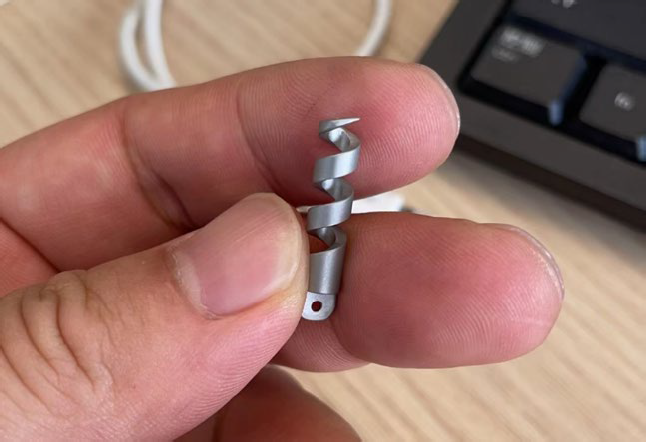
Figure 1. Specially designed hysteroscopic myoma drill.
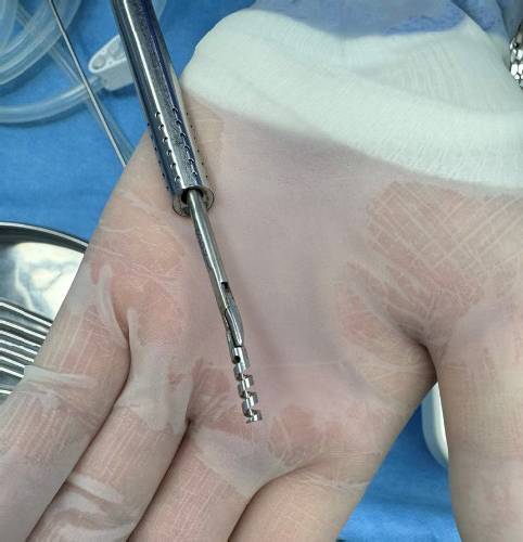
Figure 2. A 3mm needle holder grasping the myoma drill via the cold-knife hysteroscope.
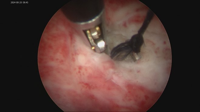
Figure 3. Anchoring and traction of the leiomyoma using the myoma drill.
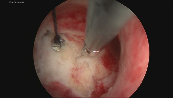
Figure 4. Dissection of the leiomyoma with scissors or forceps following traction.


