Authors / metadata
DOI: 10.36205/trocar6.vid25007
Abstract
Uterine rupture is a rare life-threatening obstetric emergency associated with poor fetal and maternal outcome, hence needs emergency surgery and prompt management. It is usually associated in cases that have history of trauma, prolonged induction or augmentation of labor, obstructed labor. Surgical history predisposing to uterine rupture includes caesarean section, myomectomy, hysteroscopy with unrecognized uterine perforation, or history of abortions. In the presented case the patient was 34 weeks pregnant, G2P1, Previous obstetrical history: full term normal vaginal birth, no previous surgeries, no medical disorders. She was not in labor. On arrival she was in constant pain and giddiness without any episode of per vaginal bleeding, with absent fetal heart sounds. On vaginal examination the cervical os was closed with no effacement.
Though the first impression was HELPP Syndrome as the ultrasound examination (US) showed ascites but in view of normal blood pressure (BP) and tachycardia a suspicion of hemoperitoneum or unrevealed injury was thought of and a computed tomography (CT) was asked for. A provisional diagnosis of uterine rupture was made as the CT showed irregular uterine wall margins with extravasation of contrast dye and hemoperitoneum, associated with a drastic fall in hemoglobin levels. She was immediately shifted to operation theatre, laparotomy was performed, uterine defect sutured, blood and blood products transfused intra-op, successfully preserving her uterus.
Case report
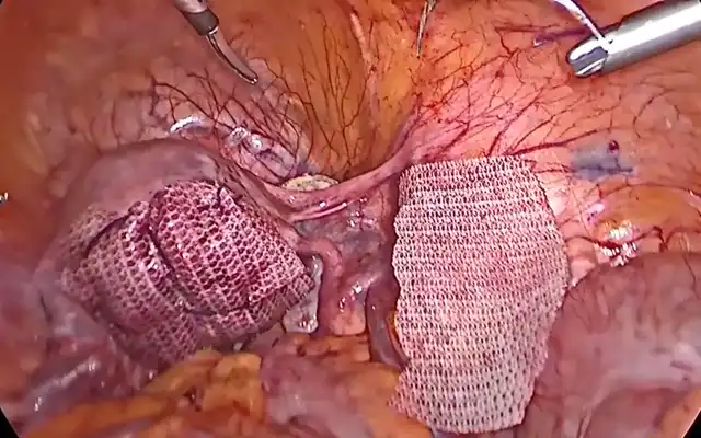
Mrs. A P, 24 yrs old lady 2nd gravida, resident of Satara, who was in her 34th. week of pregnancy with obstetric history of singleton vaginal delivery three years back and no other past surgical history (unscarred uterus) was admitted with acute onset of abdominal pain and multiple vomiting episodes in the emergency ward. Initially she was admitted in local hospital with similar complaints, managed conservatively, and then referred to our hospital for better management. On admission she had moderate pallor, blood pressure (BP) 98/60 and Pulse 154/min. On examination the uterus was mildly tense and per vaginal examination closed os. Sonography was done, that revealed an Intra Uterine Fetal Death (IUFD) along with moderate ascites and hetero-echoic collection along left anterolateral upper abdominal wall suggestive of hematoma. A day prior, a sonography was. done at outside the hospital, revealed normal fetal parameters for 34 weeks of pregnancy. Though it was a confusing diagnosis as the first thing to come to mind was an HELPP Syndrome, but she had tachycardia and the BP was normal with normal Liver function tests. This raised a suspicion of active internal bleeding and hence a CT scan of the abdomen and the pelvis was done urgently, the report did reveal an irregular left lateral wall of the uterus with extravasation of I.V contrast, with significant hemoperitoneum, raising the suspicion of a ruptured uterus. There was also a significant drop of Hemoglobin from 8.4 (a day prior) to 5.5 (on admission). Hence after proper counselling and taking due consent for a probable need to perform a hysterectomy, an emergency laparotomy was performed under General Anesthesia. The Hemoperitoneum was confirmed, around 2000 ml blood was suctioned out. A fresh stillborn male fetus of 2.1 kg was delivered by transverse incision Cesarean Section (LSCS). The uterus was found to have a large tear involving all three layers along the left posterolateral wall with irregular margins. 10×10 cm large blood clot was seen posterior to the tear. Few laceration marks were also seen along the posterior wall. Two-pints of Packed Cell Volume (PCV) were transfused intraoperatively. The ruptured site was sutured by vicryl 1-0 in 2 layers, along with the LSCS incision site. The uterus was well contracted and could be preserved. Post operatively the patient was vitally stable, monitored in ward, discharged on Post Operative Day (POD) five. Her follow up period was also uneventful.
Video clip demonstrating the amount of blood loss and active bleeding from the post lateral wall leading to a large blood clot in the contained post retroperitoneal space and hemoperitoneum.
Discussion
Uterine rupture in an unscarred uterus is highly unexpected and thus often the diagnosis gets delayed and missed at times. The patient is considered a low-risk case and hemorrhage is unrevealed since blood gets collected intra-peritoneally through the tear. The condition is usually associated with cases that have a history of trauma, prolonged induction or stimulation of labor, or over stretching of uterine wall in obstructed labor, contracted pelvis or in malpresentations. Surgical history predisposing to uterine rupture includes CS, myomectomy, hysteroscopy with unrecognized uterine perforation, a history of abortions followed by Dilatation and Curettage (D&C) or a history of instrumental vaginal deliveries in the past, to enumerate a few. In the case presented no such associated factors could be identified, thus giving us a false sense of security. However prompt actions were initiated by the team, saving the mother’s life and conserving the uterus. The patient was discharged on POD five in a vitally stable condition. Her follow up period was uneventful.
Conclusion
A pregnancy following a CS or myomectomy gets a better care and surveillance as compared to that after an uneventful vaginal birth. The rupture of an unscarred uterus causes significantly more maternal and neonatal morbidity than the rupture of a scarred uterus. Most ruptures involving unscarred uteri can be traced to one of the following etiologies: (1) trauma, (2) a genetic disorder associated with uterine wall weakness, (3) a prolonged induction or augmentation of labor, or (4) overstretching of the uterine wall. However, in this case no risk factor could be suspected that could lead to such an outcome. Hence each pregnancy should be treated with utmost attention and care, any episode of acute onset of severe abdominal pain with diffuse tenderness in the abdomen or loss of perception of fetal movements should not be ignored, all these symptoms together should raise the suspicion of a concealed uterine rupture.


