Authors / metadata
DOI: 10.36205/trocar5.2025010
Abstract
Endometriosis is a gynecological condition characterized by ectopic implantation of endometrium-like tissue. While pelvic endometriosis is a common variety, in certain rare cases urinary tract involvement may be coexistent, especially in cases of deep infiltrating endometriosis. The clinical picture may vary from asymptomatic presentation to hematuria to renal failure arising from obstruction of the ureter. Here a surgical demonstration of laparoscopic ureteral reimplantation is demonstrated in a case of deep infiltrating endometriosis with silent right sided hydroureteronephrosis
Introduction
Endometriosis is a common, benign gynecological disease affecting about 10% of the females in the reproductive age group (1). Even though endometriosis foci are benign, these are characterized by an aggressive and infiltrating growth (2). Urinary tract involvement is a very rare but serious form of infiltrating endometriosis because of the risk of urinary tract obstruction and loss of renal function (3).
Case Report
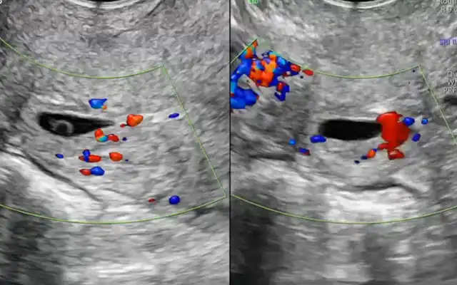
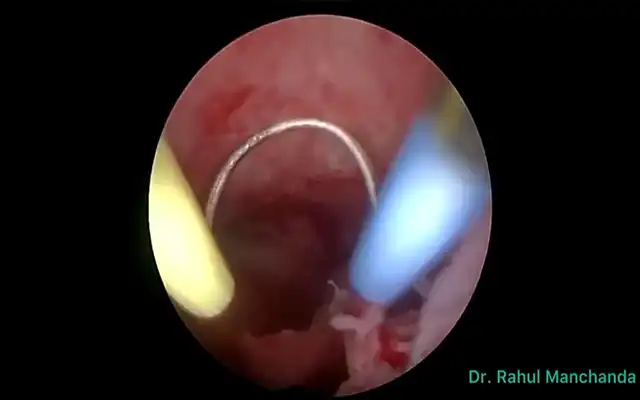
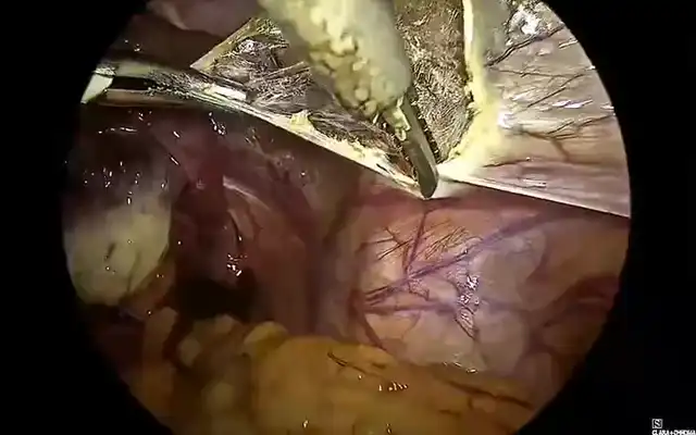
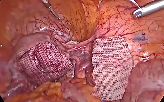
The case of a 40-year-old female with history of one caesarean section is presented. The patient had complaints of severe dysmenorrhea and chronic pelvic pain for past two years which was non-responsive to medical management, including a levonorgestrel intrauterine device. She had no bowel or bladder disturbances. Sonography incidentally detected a bladder nodule at the vesico-ureteric junction (VUJ) with gross right sided hydroureter with likely a neoplastic origin. Magnetic resonance imaging (MRI) confirmed the lesion as an endometriotic implant with a Grade 3 right sided hydroureter. Pre-operative cystourethroscopy displayed a bladder mucosal nodule in close proximity to the right ureteral orifice. The patient underwent a total laparoscopic hysterectomy with right sided salpingo-oophorectomy with excision of pelvic endometriotic implants followed by a cystotomy and excision of the nodule involving the right VUJ and terminal two cm of the ureter and the creation of a ureteroneocystostomy. Anteriorly, the nodule did obliterate the Yabuki space penetrating deep up to the bladder mucosa (as confirmed by cystoscopy). The right ureter was grossly dilated. Laterally, the nodule was in close proximity to the internal iliac artery. The ureter was dissected from its normal location at the pelvic brim up to the diseased area. The entire nodule with the involved parametrium was removed by meticulous dissection. An ureteroneocystostomy could be performed by direct ureteral reimplantation and a psoas hitch procedure was not required as a tension free anastomosed could be achieved due to the adequate mobilization of the bladder and the ureter. After four days of post-operative monitoring, the patient was discharged with a zero-pain score. At three month follow up, sonography showed resolution of hydroureter with no urinary complaints and repeat cystourethroscopy displayed a patent and functional reimplanted ureteric orifice. This is a unique case of ureteral endometriosis with the endometriotic nodule infiltrating at the right vesico-ureteric junction. It required excision of the right sided vesico-ureteric junction and terminal portion of ureter affected by intrinsic endometriosis and reconstruction of bladder with ureteroneocystostomy.
Discussion
An estimated 0.3 to 12% of women diagnosed with endometriosis also have urinary tract involvement (2). Around 50% of women with urinary tract endometriosis may be asymptomatic (4), which can lead to silent death of the kidney. Hence, it is imperative to perform sonographic monitoring and MRI guided mapping of endometriotic implants including the urinary tract. Early diagnosis and appropriate treatment will prevent complications such as hydroureter, hydronephrosis and eventual loss of renal function (5). MRI is very useful for guiding laparoscopy, and allows for the adequate evaluation of the location, size, and sub peritoneal lesion extension of deep pelvic endometriosis, providing key information for both the diagnosis and treatment planning (6). Surgical excision of the endometriotic nodules and restoration of anatomy is necessary. The surgical approach varies depending on the site and extent of bladder or ureteral involvement. Ureterolysis alone is indicated for minimal, extrinsic, non-obstructive ureteral endometriosis (7). In cases of obstructive ureteral endometriosis, surgical intervention may vary as a segmental excision and end-to-end anastomoses or a ureteroneocystostomy with or without a bladder-psoas hitch procedure or a complete uretectomy.
A pre-operative cystourethroscopy can be performed to confirm the location of a bladder nodule, if detected on sonography or MRI. If the distance between the edge of the endometriotic lesion and the inter-ureteric ridge is less than two cm, an ureteroneocystostomy is typically performed in order to reduce the risk of ureteral obstruction and fistula formation (8). An efficient surgical plan tailor-made to the patient’s diagnosis and extent of the disease with a meticulous surgical implementation can provide an optimal therapeutic outcome.
Conclusion
Endometriosis is a complex disease that challenges a gynecologist. Due to its varied presentation, we must be vigilant for urinary tract endometriosis especially in females with silent hydronephrosis. An appropriate treatment plan and the surgeon’s expertise play a pivotal role in achieving a good clinical outcome in such rare cases of infiltrating endometriosis.
Abbreviations:
VUJ- Vesico-ureteric junction
MRI- Magnetic resonance imaging


