Authors / metadata
DOI: 10.36205/trocar6.vid25011
Abstract
Objective: To describe a standardized, step-by-step surgical technique for laparoscopic small bowel resection and ileocecal valve-sparing anastomosis in deep endometriosis involving the ileum and cecum. This video aims to enhance procedural safety and reproducibility.
Introduction
Deep infiltrating endometriosis (DE) of the bowel presents a complex surgical challenge. Traditional approaches such as right hemicolectomy or ileocecal resection can lead to significant postoperative morbidity, including altered bowel function, diarrhea, and malabsorption. Involvement of the terminal ileum or cecum, particularly near the ileocecal valve, raises concerns due to the functional importance of this segment in regulating intestinal transit and absorption. Here, a standardized, valve-sparing laparoscopic technique for resection of small bowel and cecum endometriosis and with detailed intraoperative steps and outcomes, aiming to optimize disease removal while preserving function is presented.
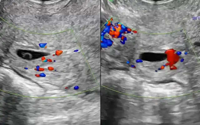
Case Presentation: A 31-year-old woman presented with severe dysmenorrhea and bowel symptoms. MRI evaluation showed: adenomyosis, posterior uterine nodule adherent to both ovaries (“kissing ovaries”), bilateral endometriomas (right ovary sum total 93 mm, left 4 mm), adhesions to the sigmoid colon with a nodular lesion measuring up to 27 mm, and serosal retraction of 10 mm. #ENZIAN classification: P0, O 1/3, T 2/2, A0, B 1/1, C0, FA, FI (sigmoid colon, 16 cm from the anorectal junction). FI: Adhesive bands reaching toward the sigmoid colon were also identified, sixteen centimeters from the anorectal junction, a long segment of bowel was found adherent to a posterior isthmic-corporeal endometriotic nodule. This segment measured up to twenty-seven millimeters in length, with a focal area of significant retraction and serosal involvement of around ten millimeters.
Intraoperatively findings instead of sigmoid lesion an ileal lesion was identified approximately some 6 cm from the ileocecal valve.
Figure 1. #ENZIAN classification map applied to the presented case of deep endometriosis. The image illustrates:
• P0: No peritoneal involvement.
• O 1/3: Right ovary with endometriomas totaling 93 mm; left ovary minimally affected (4 mm).
• T 2/2: Moderate adenomyosis.
• A0, B 1/1, C0: No anterior or rectovaginal involvement; involvement of uterosacral ligament.
• FA: Endometriotic nodule adherent to sigmoid colon at 16 cm from anorectal junction.
• FI: Ileal lesion ~10 cm proximal to ileocecal valve (not visualized on MRI but confirmed intraoperatively).
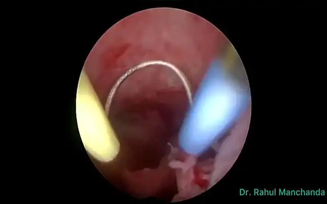
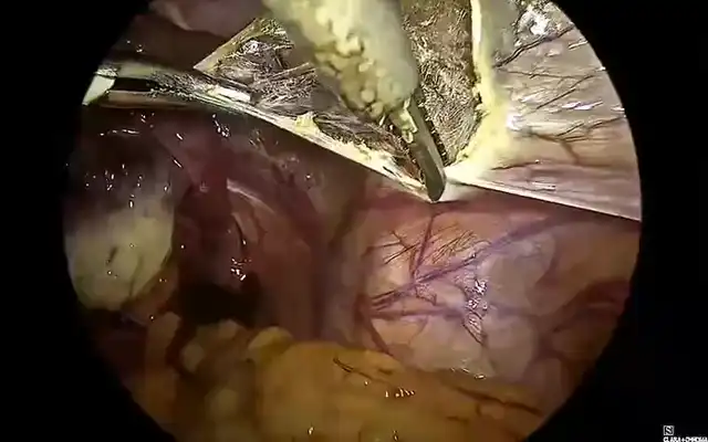
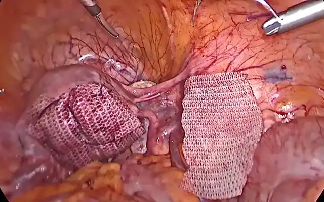
Surgical Technique: Under general anesthesia, the patient was placed in the dorsal decubitus position. Pneumoperitoneum was established via a trans umbilical incision, and five trocars were inserted using the French technique. A panoramic inspection of the abdominal cavity and pelvis was performed. All visible endometriotic lesions involving the peritoneum, ovaries, and posterior compartment were excised. For the intestinal treatment a step-by-step technique was performed:
1.Identification of the diseased ileal segment.
2.Mesenteric dissection: A minimal mesenteric window was created with the Harmonic HD 1100 ultrasonic scalpel (Ethicon), at power settings 5-7, to limit devascularization.
3.Traction suture: A suture was placed along the antimesenteric border to align the segments.
4.Enterotomies were created using the ultrasonic scalpel
5.Anastomosis: A side-to-side stapled anastomosis was performed using a 60-mm linear stapler (3.6 mm staple height), ensuring complete alignment and luminal integrity.
6.Segmental resection of the diseased ileum was completed below the enterotomies.
7.A full-thickness resection of the cecum was performed to excise a <3 cm lesion that spared the ileocecal valve and involved <1/3 of the luminal circumference
Postoperatively, the patient tolerated oral intake at 48 hours and exhibited no complications or bowel symptoms during a 6-month follow-up.
Discussion
This case illustrates a conservative and anatomically preserving approach to bowel endometriosis. Traditional ileocecal resections often result in postoperative diarrhea, bile salt malabsorption, vitamin B12 deficiency, and bacterial overgrowth due to the loss of the ileocecal valve. The valve-sparing method avoids these complications while still achieving complete lesion removal. Literature supports that excision is superior to ablation in deep lesions (ESHRE, 2022). #ENZIAN classification was used to preoperatively stage the disease; however, intraoperative findings underscored the importance of surgical mapping.
Conclusion
This video demonstrates a standardized, conservative laparoscopic technique for resection of deep endometriosis involving the ileum and cecum, highlighting the value of preserving the ileocecal valve. The method promotes functional preservation, reduces morbidity, and ensures complete excision.


