Authors / metadata
DOI: 10.36205/trocar6.vid25012
Abstract
Objective: To present a stepwise, evidence-based approach for managing severe intrauterine adhesions post-myomectomy, emphasizing the role of staged hysteroscopic adhesiolysis, mechanical barrier placement, hormonal modulation, and relook hysteroscopy to optimize clinical outcomes.
Case report: A 31-year-old nulligravida woman with primary infertility and severe intrauterine adhesions following open myomectomy and multiple failed interventions underwent hysteroscopic adhesiolysis and an inert intrauterine device was inserted as a mechanical barrier. Postoperative hormonal therapy with conjugated estrogen and medroxyprogesterone acetate was administered to promote endometrial regeneration. Second look hysteroscopy was done six weeks later. It demonstrated a restored uterine cavity and a regenerated endometrium. The patient resumed regular menstruation and reported improved clinical symptoms.
Conclusion: Severe intrauterine adhesions can be effectively managed with a stepwise protocol combining hysteroscopic adhesiolysis, mechanical barrier, hormonal therapy and relook hysteroscopy. This approach enhances uterine cavity restoration, reduces adhesion recurrence, and improves menstrual and fertility outcomes.
Introduction
Intrauterine adhesions (IUAs) result from trauma to the basal endometrium leading to fibrous bands that partially or completely obliterate the uterine cavity, causing menstrual disturbances and infertility. They may occur following dilation and curettage, myomectomy, or uterine infections, with prevalence reported as high as 19.1% depending on the extent of uterine trauma. Severe adhesions are particularly challenging to treat, as they often recur despite surgical intervention (1). Hysteroscopic adhesiolysis remains the gold standard for diagnosis and management, with outcomes improved by combining mechanical barriers, postoperative estrogen–progestin therapy, and relook hysteroscopy. Our aim in this case-based video article is to demonstrate a stepwise hysteroscopic adhesiolysis protocol, emphasizing cavity restoration with intrauterine splinting, hormonal modulation, and the importance of relook hysteroscopy in preventing recurrence (2,3).
Case report
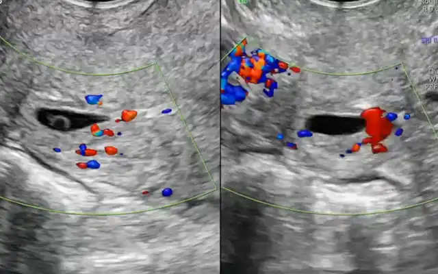
A 31-year-old nulligravida woman, working as an advocate, presented with an 8-year history of dysmenorrhea and 3–4 years of hypomenorrhea, described as scanty to moderate bleeding without clots. She had attained menarche at 13 years, with initially regular, painless cycles. Her surgical history began in 2017, when she presented with acute lower abdominal pain. Ultrasound revealed a large submucous fibroid (7.4 × 6.6 × 5.7 cm, left posterolateral wall), for which she underwent an open myomectomy. In July 2018, she developed fever, and ultrasound demonstrated hematometra with cervical stenosis; this was managed with dilation and curettage (D&C). She had been married for six years in a non-consanguineous union, with regular cohabitation but no prior pregnancies or abortions. She expressed a strong desire to conceive. In March 2019, a D&C attempted during infertility evaluation failed, as the uterine cavity could not be entered. In October 2019, she underwent diagnostic and operative hystero-laparoscopy. Findings revealed bilateral hydrosalpinx with fimbrial damage, treated by bilateral salpingectomy. Concomitant hysteroscopy documented as unhealthy endometrium (probably the false passage). Between 2021 and 2023, multiple diagnostic hysteroscopies failed. She underwent two in vitro fertilization (IVF) cycles in 2022 and 2023. Both required transabdominal trans myometrial embryo transfer due to cervical inaccessibility; however, both attempts were unsuccessful. In July 2025, pelvic ultrasound demonstrated a bulky uterus (10.1 × 8.4 × 5.7 cm) with patchy sub endometrial adenomyosis in the fundus and anterior/posterior walls, and an anterior intramural/subserosal fibroid (1.7 × 1.5 cm). A blind-ended tract (9.5 mm) extending from the endometrial cavity into the anterior myometrium, likely iatrogenic from prior trans myometrial transfers, was identified. Endometrial thickness measured 7.6 mm (done in pre-menstrual period). The cervical canal appeared tortuous, deviating leftward and turning almost 90° rightward before entering the cavity. Bilateral polycystic ovaries and a left para-ovarian cyst were also noted.
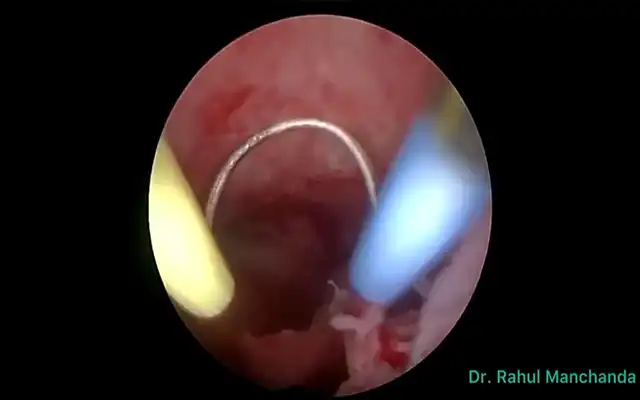
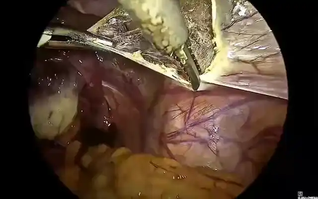
On examination, she was well nourished and vitally stable, with healed abdominal scars. Bimanual examination revealed a mobile, non-tender uterus of approximately 8-week size. The history of failed hysteroscopies and inability to enter the cavity raised a strong suspicion of intrauterine adhesions. Diagnostic hysteroscopy revealed a severely obliterated uterine cavity with dense fibrous adhesions and synechiae along the lateral walls, aligning with Grade III (Severe) as per the Manchanda’s Endoscopic Centre (MEC) classification (4). Based on the “Loddo Score: A New Intrauterine Adhesion Classification System,” the condition received a score of 14, categorizing it as moderate—indicating the need for careful management and suggesting a moderate prognosis (5). Hysteroscopic adhesiolysis was performed using cold scissors, followed by lateral wall metroplasty with a monopolar resectoscope to restore cavity anatomy. An inert intrauterine device (IUCD, copper removed) was placed as a mechanical splint. Postoperatively, the patient was given conjugated estrogen (4 mg/day for 21 days) followed by medroxyprogesterone acetate (20 mg/day for 7 days) in accordance with our unit’s standard protocol. At second-look hysteroscopy after six weeks, the uterine cavity appeared near normal, with regenerated endometrium and adequate capacity for conception. The patient resumed regular menstruation.
Video
Conclusion
Severe intrauterine adhesions represent a complex clinical challenge requiring a structured and evidence-based approach. This case demonstrates that a stepwise hysteroscopic adhesiolysis protocol—combining meticulous surgical removal of adhesions, mechanical splinting with an inert intrauterine device, and postoperative hormonal therapy—can effectively restore uterine cavity anatomy and function. The addition of relook hysteroscopy is critical in confirming successful cavity restoration and minimizing adhesion recurrence. Such comprehensive management not only improves menstrual outcomes but also optimizes the uterine environment for potential fertility restoration, providing hope beyond adhesions for affected women.