Authors / metadata
DOI: 10.36205/trocarvid2.2021001
Abstract
Study Objective: to demonstrate the laparoscopic diagnosis and management of a rare class U6 malformation, namely the accessory cavitated uterine mass (ACUM).
Design: A didactic description of the diagnostic and surgical steps through a case report.
Setting: A private hospital in Rome, Italy.
Patient: 55-year-old, gravida 0 para 0 patient, with a history of conization for a high-grade intraepithelial lesion and a xipho-pubic laparotomy for a recent car accident trauma. Few months after surgery, the patient underwent a control MRI, incidentally revealing a latero-uterine mass, with liquid content, requiring further investigation.
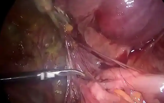
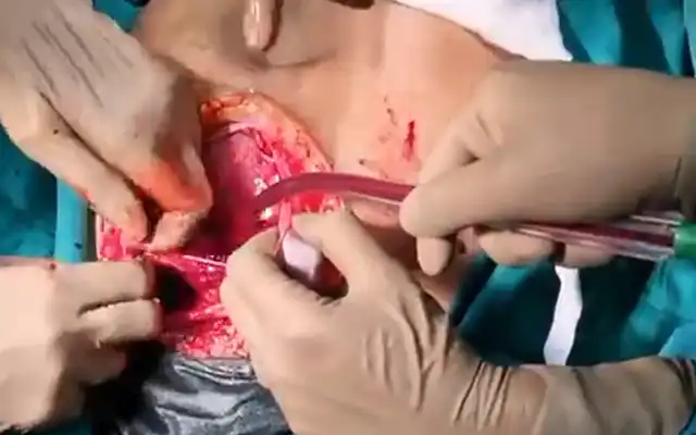
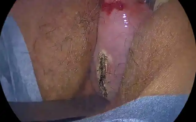
Intervention: Laparoscopy revealed a uterus deformed by a large mass situated at the right wall, under the round ligament, arousing suspicion of a non-communicating rudimentary horn with hematometra. The insertion of the ipsilateral Fallopian tube and utero-ovarian ligament indicated the right horn of a normally shaped uterus, thus orienting towards an ACUM. Incision and dissection of the mass led to the release of a chocolate fluid which was drained. The cavitated mass was completely excised.
Main result: Histological exam confirmed a uterus-like mass composed of a myometrial wall and an endometrium.
Corresponding author: Amal Drizi
Conclusion
ACUM belongs to the rare unclassified uterine malformations, poorly known by practitioners, and often misdiagnosed for a non-communicating rudimentary horn with hematometra or an adenomyotic cyst. The landmarks for a proper laparoscopic diagnosis and management are to be stressed.