Authors / metadata
DOI: 10.36205/trocar3.2021002
Abstract
Background: Cesarean scar pregnancy (CSP) is defined as the implantation of a gestational sac within the scar of a previous cesarean surgery.
Cases: Two cases of CSP treated in Mohammed IV University Hospital are reported. The first case is a deep implantation CSP managed by laparoscopy ligation of the uterine arteries was performed with minimal bleeding. The second case is a deep implantation CSP managed by laparoscopy without clamping of the uterine arteries resulting in massive hemorrhage.
Conclusion: For CSP, temporary Bilateral Uterine Artery Ligation may be a reasonable and secure procedure in the laparoscopic management of patients who had a cesarean scar pregnancy and do desire to preserve their fertility.
Introduction
Cesarean scar pregnancy (CSP) is a rare form of ectopic pregnancy however because of the increase in cesarean deliveries, it has become increasingly common over the last decades [1]. Two types are described, type I or endogenous when the implantation is in the scar with development towards the uterine cavity or towards the cervico-isthmic canal, CSP Type 2 or exogenous here the implantation occurs deep in the scar with development towards the bladder and towards the abdomen with an important risk of rupture [2].
This special type of ectopic pregnancy poses a great reproductive risk for women of childbearing age with a history of cesarean section. However, there is no consensus on the treatment of CSP. Here two cases of cesarean scar pregnancy managed by laparoscopy are presented.
Case report 1
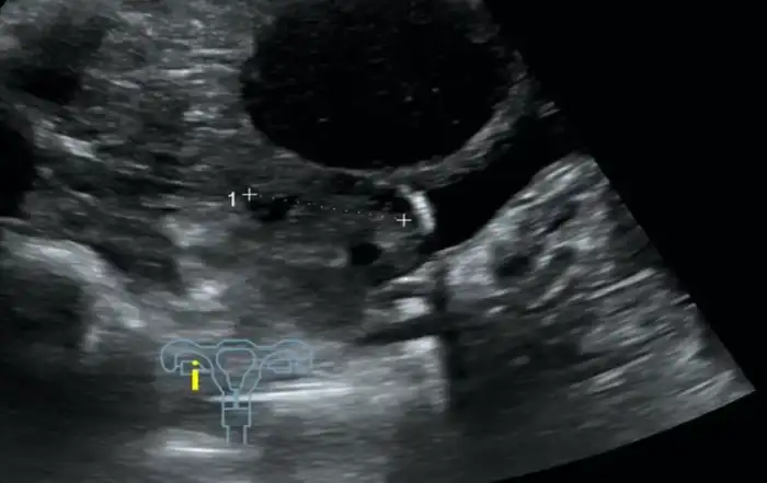
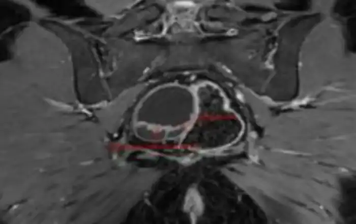
A 28-year-old woman gravida 3 para 2 with a history of 2 cesarean deliveries. the patient presented with a minimal blackish bleeding at an amenorrhea of 8 weeks. Beta-human chorionic gonadotropin (bHCG) was 53280 mIU/ml. On transvaginal ultrasonography, an 8- week fetus was identified in the cesarean scar defect (type II), an MRI revealed a gestational sac embedded in the hysterotomy scar, coming down to the serosa without interposition of the myometrium, lateral on the right side. Detailed information and treatment options were discussed with the patient, and laparoscopy was retained.
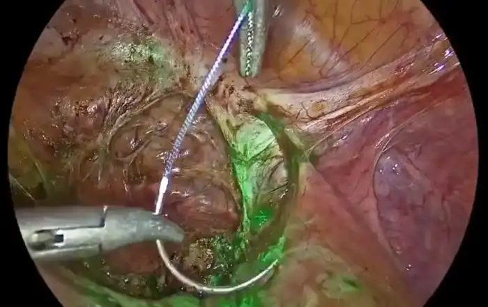
Laparoscopic and hysteroscopic exploration were performed at first reveling a dense adhesion between the omentum and the uterine surface, an adhesiolysis was performed. Bilateral uterine artery ligation was performed to minimize blood loss. The bladder was dissected down to expose the vesico-vaginal space, a protrusion from the anterior wall of the uterus located at the uterine isthmus was found. An incision over the bulge was done then the trophoblastic tissue was removed. A suction curettage was performed followed by a repair of cesarean scar dehiscence. Estimated blood loss was 200 ml. The patient was discharged two days after the surgery. Nine months later, the patient had a spontaneous singleton pregnancy.
Case report 2
A 36-year-old woman gravida 3 para 2 with a history of 2 cesarean deliveries presented at outpatients for routine examination at 6 weeks of amenorrhea with no bleeding or pain. Beta- human chorionic gonadotropin (bHCG) was 12560 mIU/ml, transvaginal ultrasound revealed a CSP (type II) with fetal cardiac activity. MRI did reveal that the placenta invaded the myometrium up to contact with the serosa, responsible for a bulging of the anterior face of the isthmic region that joins the posterior wall of the bladder without a separation border. Laparoscopic management was opted for. During the exploration, a dense adhesion was observed between the cesarean scar and the left lateral abdominal wall. After adhesiolysis, during the attempt to push down the bladder to expose the lower uterine scar, a massive hemorrhage did occur.
An aspiration of trophoblastic tissue to reduce the hemorrhage was performed before completing the dissection. the scar tissues were cut and the dehiscence was repaired. The estimated blood loss was 1200 ml. Patient was transfused with three (Author numbers under ten are best written in letters) units of red blood cells. T h e patient was discharged 3 days later with 10 g/dl hemoglobin.
Discussion
Obviously, laparoscopic management is not the only way to manage a CSP, it could be managed by medical treatment or by hysteroscopy and vacuum aspiration. According to the latest Expert Consensus on the Diagnosis and Treatment for Cesarean Scar Pregnancy laparoscopic surgical treatment is recommended in CSP type II [3].
The purpose of presenting those two case reports, is to draw attention on the importance of bilateral uterine artery ligation UAL in the laparoscopic management of CSP.
There have been several studies concerning the use of temporary UAL in the treatment of CSP, however no cases of failed therapy or complication have been documented. Chen et Al [4] conducted a study of the clinical efficacy of Temporary UAL during laparoscopy in the treatment of CSP on 83 patients it shows that the UAL patients did better then the patients in the no ligation group. Also, Huang et Al [5] demonstrated in a retrospective comparative study of 173 patients with CSP that clinical outcomes were better with hysteroscopic and laparoscopic surgery using temporary UAL than other surgical management techniques.
In other randomized controlled trial studies permanent or temporary UAL have been showed to be effective strategies to reduce blood loss in surgeries implying incision and repair of the myometrium [5,6].
UA embolization could be a good alternative as a pre-treatment for decreasing the risk of bleeding. In a systematic review concerning the efficacy and safety of treatment options, UAE was associated with a success rate of 93%, a risk of hemorrhage of 5%, and a risk of hysterectomy of 3% [7]. However, post- embolization syndrome, pelvic infection, damage to the ovarian function, or endometrial atrophy leading to amenorrhea, intrauterine adhesions, and infertility could occur, affecting the reproductive function of patients.
In conclusion laparoscopic temporary UAL reveals itself to be a better choice and a safer procedure for the treatment of CSP mainly in type II especially for patients with a desire for future fertility.
Abbreviation
CSP: cesarean scar pregnancy
bHCG: beta Human chorionic gonadotropin
Conclusion
The hysteroscopic view of cystic atrophy correlates 60 % with histopathology in the preliminary analysis. Cystic atrophy does not increase the risk of endometrial carcinoma, but presence of metabolic diseases may pose a risk of endometrial cancer due to the increased estradiol levels.
Hysteroscopic view: cystic atrophy. Immunohistochemistry may add value if the density of estrogen and progesterone receptors in the epithelial cells is high, in women with metabolic diseases and under evaluation for postmenopausal bleeding (9).
Limitations: The amount of endometrial tissue available for HPE varies in the two techniques of targeted biopsy and visual dilatation and curettage. Targeted biopsy with resection of the area or visual dilatation and curettage may increase the accuracy of the hysteroscopic view.
Strength: This is a prospective cohort ongoing study with a follow up of 12 months and aim to follow for 10 years.
Learning points: Evaluation for postmenopausal women must include a transvaginal ultrasound, hysteroscopy, as the technique involves direct vision, and an endometrial biopsy. Performing serum concentration of E2 may help in the management plan.