Information
- Author: Alfonso Rossetti / Ornella Sizzi
A 36 years old patient 0 para was referred to our Department for the presence of two myomas: a 7 cm anterior myoma and a 5 cm lateral infralegamentous myoma. The Sonographic preoperative evaluation showed irregular anaechogenic area like for colliquative necrosis The level of LDH isoenzyme 3 was 23.
Surgical procedure
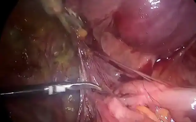
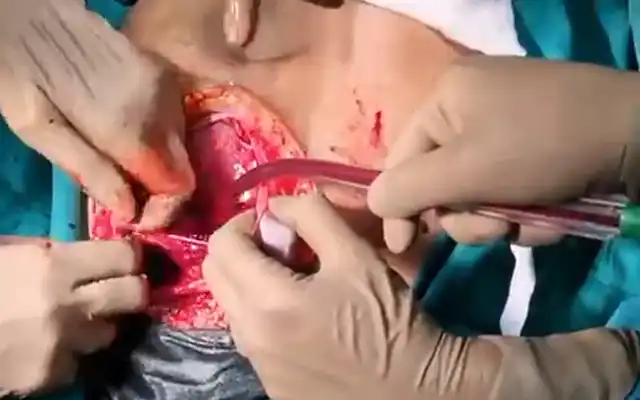
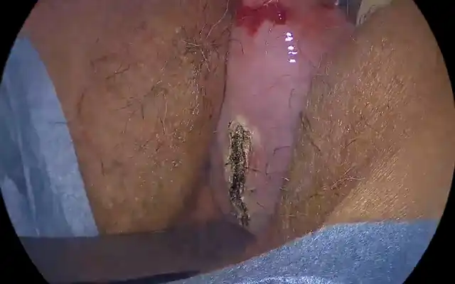
The myometrium surrounding the myoma was injected with a diluted solution of vasopressin (20 UI in 400 ml of saline). A longitudinal incision was performed on the anterior wall of the uterus. The myoma capsule was then opened. The myoma was very soft and filled with fluid. Therefore, the myomectomy was performed with the stripping technique usually used for the ovarian cysts. The lateral myoma was also colliquated and came out together with the anterior one. The suture was then performed with figure of eight introflecting stitches. The myoma was not morcellated but was extracted from the abdominal cavity using an endobag.The Post-Operative Course was uneventful and the patient was discharged in the second postoperative day. The Histologic examination confirmed the diagnosis of uterine myoma with colliquative necrosis.
Video
MEMBERS AREA
Categories